
Worried about unexpected vet bills?
Pet insurance can cover thousands in unexpected vet costs. Get a free quote from Lemonade in under 2 minutes.
Get My Free Quote →Sponsored · Opens Lemonade.com
Brain regeneration represents one of nature’s most remarkable abilities. While humans cannot naturally regrow brain tissue after injury, numerous creatures across the animal kingdom possess this extraordinary capacity. From simple invertebrates to more complex vertebrates, these animals challenge our understanding of neural regeneration and offer insights that might someday lead to revolutionary treatments for human brain injuries and neurodegenerative diseases. This exploration of 13 remarkable creatures highlights the diverse approaches to brain regeneration found throughout nature, demonstrating the resilience and adaptability of life on Earth.
The Science Behind Brain Regeneration

Brain regeneration involves the restoration of neural tissue after damage through either replacement of damaged neurons or growth of new neurons (neurogenesis). In most mammals, including humans, neurogenesis is extremely limited in adulthood, occurring primarily in the hippocampus and subventricular zone. However, many other animals possess neural stem cells that remain active throughout life, allowing them to repair damaged brain tissue. These animals typically maintain pools of undifferentiated cells that can proliferate and differentiate into neurons when needed. The mechanisms behind this remarkable ability vary widely across species, involving different signaling pathways, gene expression patterns, and cellular responses to injury. Understanding these mechanisms provides valuable insights for regenerative medicine and potential treatments for human neurological conditions.
Planarians Masters of Whole-Brain Regeneration
Planarians, flatworms of the phylum Platyhelminthes, demonstrate perhaps the most impressive brain regeneration capabilities in the animal kingdom. These small, aquatic creatures can regenerate their entire bodies, including their brains, from fragments as small as 1/279th of the original animal. The secret to this extraordinary ability lies in their abundant pluripotent stem cells called neoblasts, which make up roughly 30% of all planarian cells. When a planarian is cut, neoblasts migrate to the wound site and differentiate into the required cell types, including neurons. Remarkably, the regenerated brain not only develops the correct structure but also retains memories from before the injury. This indicates that memory in planarians may be stored throughout the body or that the regeneration process involves precise replication of neural connections. Scientists studying planarians have identified key genes and signaling pathways involved in brain regeneration, including the Wnt/β-catenin pathway, which could inform future approaches to treating human brain injuries.
Sea Cucumbers Neural Regeneration Through Evisceration

Sea cucumbers, marine invertebrates of the class Holothuroidea, employ a dramatic defense mechanism called evisceration that showcases their regenerative abilities. When threatened, certain species expel their internal organs, including parts of their nervous system, only to regrow them later. Their nervous system consists of a ring around the mouth and five radial nerves, which they can substantially regenerate after injury or evisceration. This process takes about 3-5 weeks and involves dedifferentiation of existing cells, proliferation, and redifferentiation into neural tissue. Sea cucumbers produce specific neurotrophic factors that guide this regeneration, ensuring proper neural connections are established. Scientists have identified several bioactive compounds in sea cucumber tissues that promote cell growth and healing, including glycosides, peptides, and polysaccharides. These compounds have potential applications in developing therapies for human neurological disorders. The sea cucumber’s regenerative capabilities highlight nature’s ingenious solutions to survival challenges and offer valuable models for studying neural regeneration.
Axolotls The Regeneration Champions
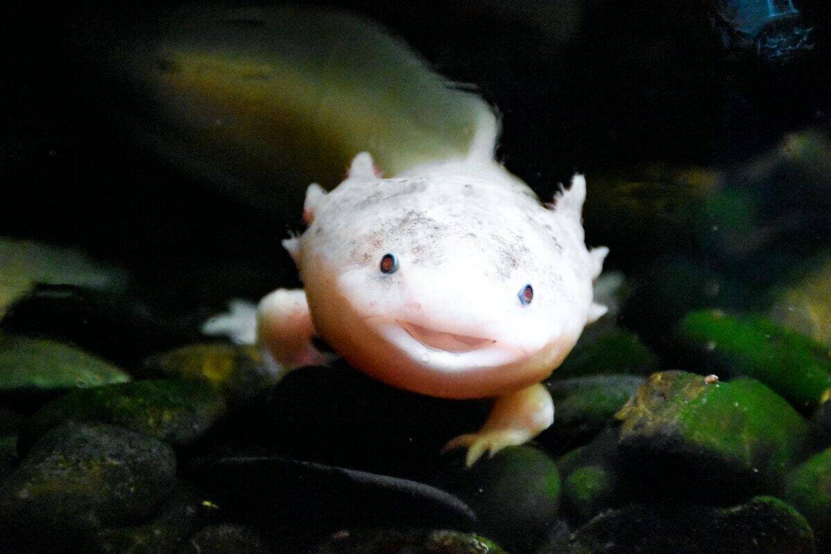
Axolotls (Ambystoma mexicanum), aquatic salamanders native to Mexico, are renowned for their extraordinary regenerative abilities, which extend to portions of their brains. These neotenic amphibians can regenerate not only limbs, tail, and heart tissue but also significant parts of their central nervous system, including areas of the brain. Unlike most vertebrates, axolotls maintain neural stem cells throughout adulthood, particularly in the telencephalon. After brain injury, these stem cells proliferate rapidly, migrating to damaged areas and differentiating into appropriate neural cell types. The regeneration process involves complex molecular signaling, including factors like fibroblast growth factor (FGF) and sonic hedgehog (Shh). Remarkably, axolotls can regenerate up to 70% of the cerebral hemisphere volume within 12 weeks following injury. This regeneration is functionally significant, with studies showing recovery of cognitive abilities and behaviors controlled by the damaged brain regions. The axolotl genome, which is ten times larger than the human genome, contains unique genetic elements that may explain these exceptional regenerative capabilities. Scientists have identified several genes specifically upregulated during brain regeneration in axolotls that are inactive or absent in mammals, providing potential targets for therapeutic development.
Zebrafish The Model Organism for Brain Regeneration

Zebrafish (Danio rerio) have become invaluable model organisms for studying brain regeneration due to their remarkable neural plasticity and experimental advantages. Unlike mammals, adult zebrafish can regenerate various regions of their brains following injury through robust neurogenesis. This process is particularly evident in the telencephalon, where radial glia cells act as neural stem cells, proliferating to replace damaged neurons. Following traumatic brain injury, zebrafish activate specific regeneration programs within 24 hours, with new neurons integrating into existing circuits within 4-8 weeks. The regenerative response includes inflammation control, activation of developmental signaling pathways, and epigenetic reprogramming. Zebrafish brains contain approximately 16 distinct neurogenic niches compared to just two in mammals, explaining their superior regenerative capacity. Their transparent embryos and relatively simple brain structure, containing about 100,000 neurons (compared to 86 billion in humans), allow researchers to directly observe neural regeneration processes. Studies of zebrafish have identified key molecules involved in brain regeneration, including cystatin F, chemokine signals, and fibroblast growth factors, providing potential therapeutic targets for human neurological conditions. The genetic similarity between zebrafish and humans—they share about 70% of their genes—makes findings from these studies particularly relevant to human medicine.
Crayfish Growing New Neurons from Blood Cells

Crayfish exhibit a unique approach to brain regeneration that involves transforming blood cells directly into neurons. These freshwater crustaceans possess a neurogenic niche in their brains containing neural precursor cells that continuously produce new neurons throughout their lives. What makes crayfish remarkable is that these precursor cells appear to be replenished by hemocytes (blood cells) that migrate into the brain and transform into neural stem cells. This process is particularly active in response to environmental enrichment or brain injury. Studies have shown that crayfish can generate thousands of new neurons daily in certain brain regions, particularly those responsible for learning, memory, and sensory processing. The transformation of hemocytes into neurons involves a complex sequence of cellular reprogramming controlled by specific transcription factors and signaling molecules. This mechanism differs dramatically from that seen in most other animals and suggests an evolutionary adaptation to maintain cognitive function throughout the crayfish’s life. The discovery of this blood-to-brain transformation pathway in 2014 by researchers at the University of Kentucky opened new perspectives for potential regenerative therapies, as it demonstrates a natural example of cellular transdifferentiation—the conversion of one specialized cell type directly into another without reverting to a stem cell state.
Sea Stars Regenerating Complex Neural Networks

Sea stars (starfish) demonstrate remarkable neural regeneration capabilities as part of their broader regenerative abilities. When a sea star loses an arm, it must regenerate not only the limb but also the complex radial nerve that controls it. This radial nerve is an extension of the sea star’s decentralized nervous system, which consists of a central nerve ring and radial nerves extending into each arm. The regeneration process begins with wound healing and the formation of a blastema—a mass of undifferentiated cells that will develop into the new arm and its associated neural structures. Within this blastema, specialized cells called radial glia act as neural progenitors, proliferating and differentiating to form new neurons and supporting cells. The regeneration follows a proximal-to-distal pattern, with neural tissue closest to the central disc forming first. Complete regeneration of an arm and its neural components typically takes 3-12 months, depending on the species and environmental conditions. Remarkably, the regenerated neural tissue forms appropriate connections with the existing nervous system, restoring functional control of the new arm. Recent studies have identified several signaling molecules crucial for this process, including bone morphogenetic proteins (BMPs) and transforming growth factor-beta (TGF-β), which coordinate the timing and patterning of neural regeneration.
Spiny Mice Mammalian Brain Regeneration Pioneers

Spiny mice (Acomys species) represent a rare example of mammals with enhanced regenerative capabilities, including limited brain regeneration. Native to Africa and the Middle East, these rodents are known for their ability to shed and regrow skin without scarring, regenerate hair follicles, sweat glands, cartilage, and even portions of their spinal cord. Recent research has revealed that spiny mice also show enhanced neural regeneration compared to laboratory mice. Following traumatic brain injury, spiny mice exhibit reduced glial scarring and increased neurogenesis in the hippocampus and surrounding damaged areas. They maintain a larger pool of neural progenitor cells into adulthood and show upregulation of growth factors that promote neural survival and regeneration. The inflammatory response in spiny mice brains also differs from that of other mammals, favoring an M2 anti-inflammatory macrophage response rather than the pro-inflammatory M1 response typical in non-regenerative species. This creates a more permissive environment for neural repair. Studies have shown that spiny mice can regenerate approximately 10-15% more neurons after brain injury compared to laboratory mice, with corresponding improvements in cognitive recovery. The genetic and molecular mechanisms underlying these enhanced capabilities are being intensely studied, as they may represent an evolutionary intermediate that could provide insights into how to enhance regeneration in other mammals, including humans.
Hydra Immortal Regenerators
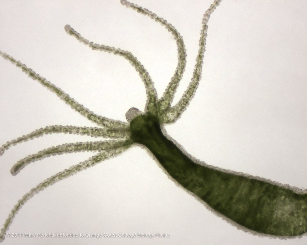
Hydra, tiny freshwater cnidarians, possess perhaps the most extreme regenerative capabilities in the animal kingdom, including complete brain regeneration. These simple organisms, measuring just 0.5-1.5 cm in length, can regenerate their entire body from small fragments representing as little as 1/200th of the original animal. Their nervous system consists of a diffuse nerve net with a concentration of neurons forming a rudimentary brain-like structure near the mouth called the circumoral nerve ring. When decapitated, hydra can regenerate this neural structure within 72 hours through a process that involves the activation of epithelial stem cells. These cells proliferate and differentiate into new neurons guided by Wnt and bone morphogenetic protein (BMP) signaling pathways. Unlike most animals, hydra maintains three distinct stem cell populations throughout its body that continuously replace all cell types, contributing to both regeneration and potentially unlimited lifespan. These stem cells respond to injury by increasing proliferation rates and altering their differentiation patterns. Research has shown that hydra regeneration depends on precise bioelectrical signals that establish body polarity and guide the regeneration process. The genes involved in hydra neural regeneration show remarkable conservation with those in higher animals, including humans, suggesting ancient evolutionary origins for regenerative mechanisms. This genetic conservation makes hydra valuable models for understanding fundamental aspects of neural development and regeneration applicable across species.
Octopuses Regenerating Complex Neural Circuits
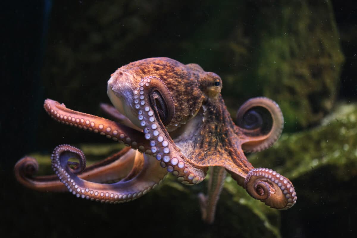
Octopuses demonstrate remarkable neural regeneration capabilities within their complex nervous system. These cephalopods possess a distributed nervous system with approximately 500 million neurons—comparable to that of a dog—with two-thirds located in their arms rather than their central brain. When an octopus loses an arm, it can regenerate not only the limb but also the complex neural circuits within it. Each arm contains about 40 million neurons organized into ganglia that control movement, sensation, and even learning, functioning with significant autonomy from the central brain. The regeneration process begins with wound healing followed by blastema formation. Neural progenitor cells proliferate within this blastema, differentiating into various neuronal types and forming new ganglia. The regenerated neural tissue establishes appropriate connections with both local structures and the central nervous system. Complete neural regeneration in an arm typically takes 3-6 months, with functional recovery occurring gradually during this period. Studies have identified several neurotrophic factors and transcription factors uniquely expressed during octopus neural regeneration, including octopus-specific variants of genes like Pax6 and Notch, which are conserved across animal phyla. Research has shown that octopuses can maintain memories and learned behaviors after partial brain injury, suggesting effective neural repair mechanisms or compensatory plasticity in their central brain. This combination of a complex nervous system with significant regenerative capacity makes octopuses particularly valuable models for understanding neural repair in advanced brains.
Newts Life-Long Brain Regenerators

Newts stand out among vertebrates for their extraordinary ability to regenerate brain tissue throughout their entire lifespan, even in old age. These amphibians can regenerate various parts of their central nervous system, including substantial portions of their brain, retina, and spinal cord. Following brain injury, newts initiate a complex regeneration process that begins with the rapid formation of a blood clot and activation of immune responses. Unlike mammals, whose immune response often inhibits regeneration, the newt’s immune system promotes it by clearing debris and releasing growth factors. The key to newt brain regeneration lies in their ability to reprogram mature cells through a process called dedifferentiation. Resident ependymoglial cells lining the ventricles of the brain can dedifferentiate, proliferate, and then redifferentiate into new neurons and glial cells as needed. This process involves the temporary suppression of tumor suppressor proteins like p53, allowing cells to re-enter the cell cycle. Studies have shown that newts can regenerate approximately 50% of their dopaminergic neurons in the midbrain within 30 days after ablation—an ability particularly relevant to Parkinson’s disease research. The regenerated neurons successfully integrate into existing neural circuits, restoring lost functions. Recent genomic analyses have revealed that newts possess unique gene expression patterns and specialized variants of proteins involved in extracellular matrix remodeling and cell cycle regulation that may facilitate their exceptional regenerative abilities.
Lizards Beyond Tail Regeneration

While lizards are famously known for tail regeneration, certain species also demonstrate limited brain regeneration capabilities. The regenerative abilities vary significantly across the approximately 6,000 lizard species, with some showing more extensive neural repair than others. The leopard gecko (Eublepharis macularius) has been particularly well-studied, showing notable neurogenesis in specific brain regions after injury. Following traumatic brain injury, these geckos activate neural stem cells in the ventricular zone, which proliferate and migrate to damaged areas. This process involves upregulation of developmental genes including Sox2, Pax6, and Nestin, which are typically inactive in adult mammalian brains. The medial cortex of lizards, which is homologous to the mammalian hippocampus, shows especially robust regenerative capacity, with new neurons integrating into existing circuits within 2-4 weeks after injury. Research has identified several unique aspects of the lizard inflammatory response that may facilitate brain regeneration, including specialized macrophage activation patterns that limit secondary damage and promote tissue repair. Interestingly, lizard species that can change color, such as chameleons, show enhanced neural plasticity and regeneration in brain regions controlling this ability. Studies comparing closely related lizard species with different regenerative capacities have identified genetic and environmental factors that influence neural repair, providing valuable comparative models. The seasonal variation in lizard brain regeneration, with enhanced capabilities during warmer months, suggests metabolic and hormonal influences on neural repair mechanisms that may have relevance for therapeutic approaches.
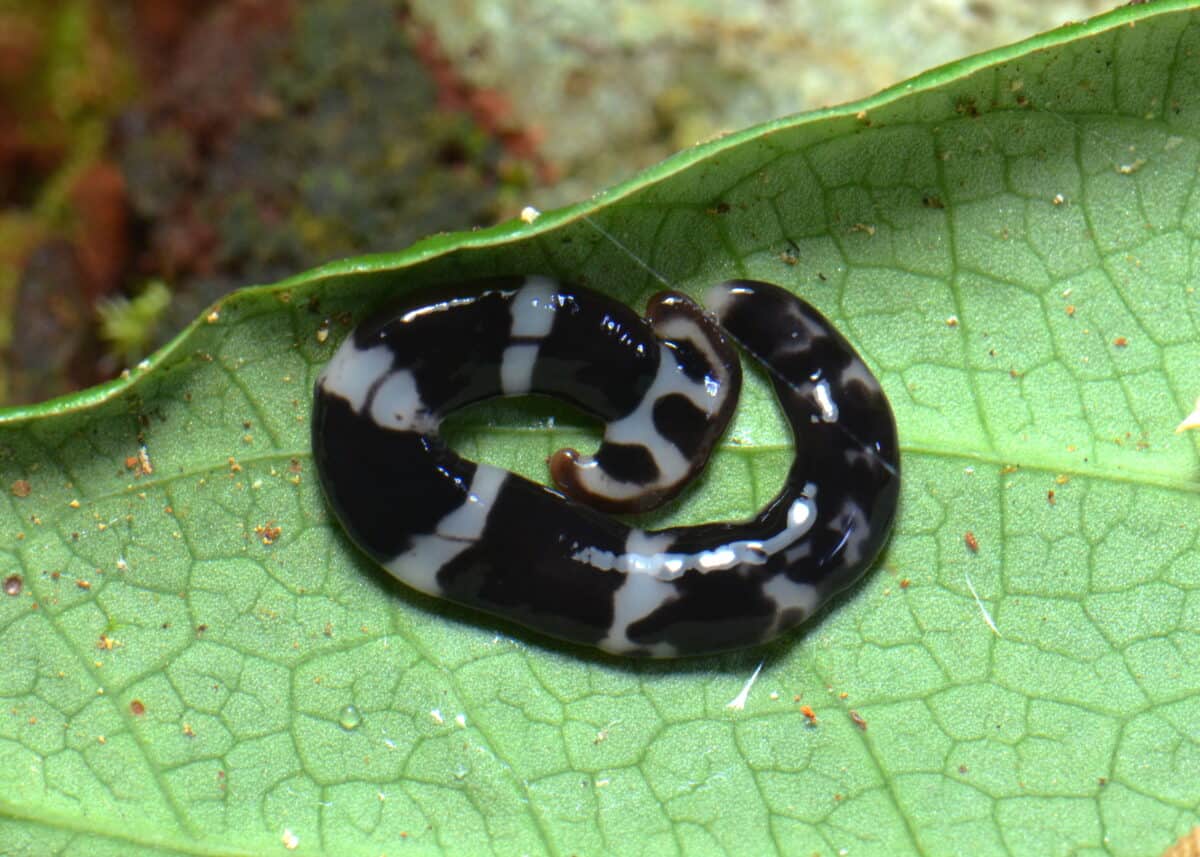
Leeches Simple Yet Effective Neural Regeneration
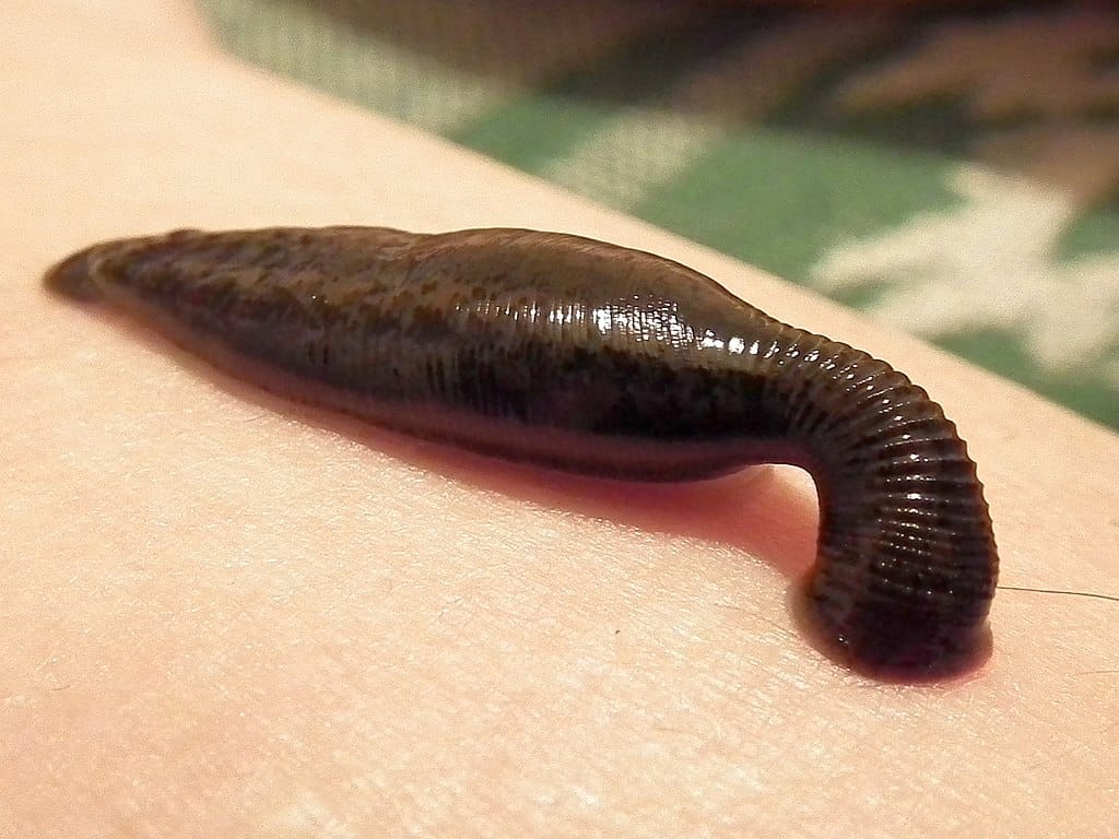
Leeches exhibit remarkable neural regeneration capabilities despite their relatively simple nervous system. These annelids possess a rope-ladder-like nervous system consisting of a chain of ganglia connected by nerve cords, with approximately 10,000 neurons organized into 21-34 segmental ganglia, depending on the species. When a leech’s nervous system is damaged, it initiates a well-coordinated regeneration process. Within hours of injury, microglial cells (the leech equivalent of immune cells in the nervous system) migrate to the damage site to clear debris and release factors that stimulate repair. Severed axons can regenerate and reconnect with their original targets with surprising precision, restoring neural function.
Conclusion

The ability to regrow brain tissue is not confined to science fiction—it’s a biological reality for a surprising range of animals. From simple invertebrates like hydra and planarians to more complex vertebrates like axolotls and newts, these 13 creatures showcase nature’s astonishing regenerative toolkit. Their diverse mechanisms—from stem cell proliferation to transdifferentiation—offer powerful models for scientific exploration and medical innovation. As researchers continue to decode the genetic and cellular secrets behind brain regeneration, these animals could hold the key to unlocking future therapies for brain injuries, stroke, and neurodegenerative diseases in humans. The study of these species not only deepens our appreciation of biological resilience but also brings us closer to the possibility of healing the human brain.
Worried about unexpected vet bills?
Pet insurance can cover thousands in unexpected vet costs. Get a free quote from Lemonade in under 2 minutes.
Get My Free Quote →Sponsored · Opens Lemonade.com
As a little kid, I fell in love with nature, wildlife, and animals. Living in the USA, South Africa, Italy, China and Germany gave me the opportunity to discover the world's Wildlife. My favorite animals are Mountain Gorillas, Siberian Tigers, and Great White Sharks.
I'm a certified PADI Open Water Diver, went to Everest Base Camp and Trekked Gorillas in Uganda. I hold a Master of Science in Economics and Finance.
Please send any feedback to feedback@animalsaroundtheglobe.com
- 5 Animals That Can Travel Thousands of Miles Without Stopping - June 6, 2026
- 12 Fish That Hunt in Packs Like Wolves - June 6, 2026
- The Vibrant Danger of Poison Dart Frogs - June 6, 2026
Leave a comment
You must be logged in to post a comment.